
Propelling diagnostic innovation
Getica AB – A part of Gentian Diagnostics ASA
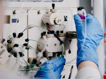
Innovation of new immunodiagnostic assays
Getica is a biotechnological company located at the Sahlgrenska Science Park in Göteborg, Sweden. Our core activities have since 2014 been centered around antigens and antibodies, of both avian and mammalian origin, for research and industrial use in various diagnostic assays. In 2023, Getica was acquired by Gentian Diagnostics, and since then Getica has put greater focus at R&D activities. Together with Gentian, our primary focus and aim is to put new and innovative immunodiagnostic tests on the global IVD market.

Contact details
Mail: info@getica.se
Telephone: +4672-858 81 87
Getica AB
Medicinaregatan 9C
413 90 Göteborg
Org: 556615-6112